Figure 2.

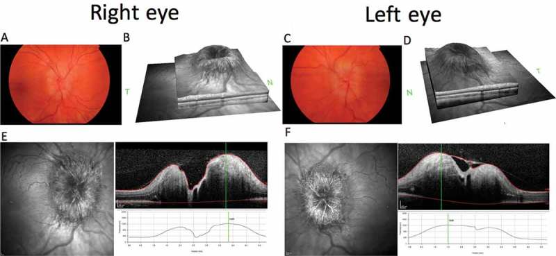
Composite figure showing colour fundus photographs of the papilloedema affecting the optic nerve head (OHN) of the right (A) and left (C) eyes. Optical coherence tomography (OCT) SPECTRALIS HRA+OCT (Heidelberg Engineering, Heidelberg, Germany), infrared (IR) images of the ONH, and volume cross-sectional images and the elevated height through the centre of the ONH of the right (E) and left (F) eyes. OCT IR disc volume reconstructions for right (B) and left (D) eyes.
