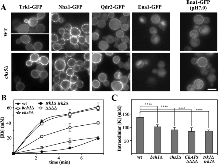
FIGURE 2:
The localization of PM transporters in exomer mutants. (A) Localization of different PM transporters in wild-type and chs5∆ strains. Proteins were chromosomally appended with the GFP at their C-terminus, except Trk1-GFP, which was expressed from plasmid pRS414. All proteins were visualized in cells growing in nonbuffered SD media except Ena1-GFP, which was also visualized at pH 7.0. Note the similar localization in wild-type and chs5∆ strains. (B) Rubidium uptake in the different mutants and (C) intracellular levels of potassium. The strain labeled ∆∆∆∆ corresponds to strain YAS563-16a in which all four ChAPs have been deleted (see Table 1).

