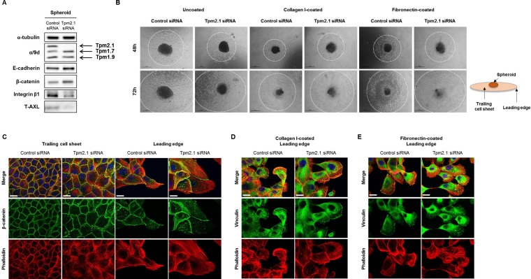
Figure 8. Rate of migration from spheroid depends on microenvironment.
(A) RNAi treated MCF10A cells were re-cultured in non-adherent plate and protein expression of total cell lysates was detected after 96 hours. (B) Spheroids were re-cultured on different substrates coated on culture plates to measure cells migrating out of the spheroids at 48 and 72 hours (Scale bar: 500 μm). (C) Immunofluorescent data for β-catenin, phalloidin and DAPI were imaged at the trailing cell sheet and leading edge of spheroid migration under uncoated culture condition. (D–E) Cells migrating out of the spheroid on collagen I and fibronectin were stained for vinculin, phalloidin and DAPI (Scale bar: 20 μm).

