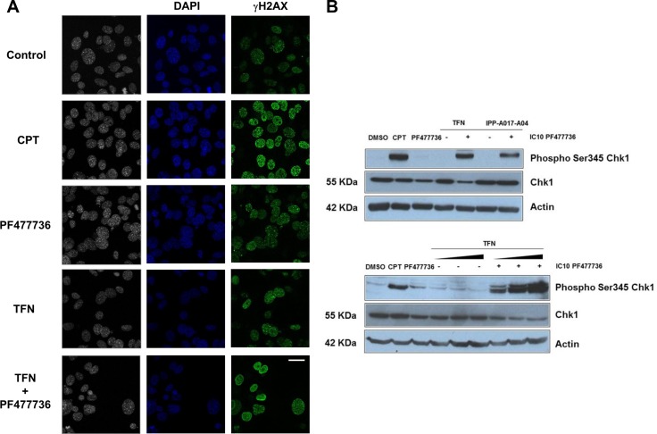
Figure 5. The combination of teriflunomide and PF477736 increases the amount of DNA damage in transformed MEFs.
(A) Immunofluorescence of DNA (blue) and γH2AX (H2AX phosphorylation on serine 139) (green) in transformed MEFs that were exposed to either IC70 TFN (10 μM), IC10 PF477736 (0.7 μM), the combination of these compounds or 0.1 μM positive control camptothecin (CPT). Scale bar, 20 μm. (B) Western blotting analysis of Chk1 phosphorylation on serine 345 in cell lysates prepared 8 hours after the beginning of the exposure. Upper panel: transformed MEFs were exposed to vehicle, 0.1 μM camptothecin, IC10 PF477736, IC70 TFN ± IC10 PF477736 and IC70 IPP-A017-A04 (22 μM) ± IC10 PF477736. Lower panel: cells were exposed to vehicle, 0.1 μM camptothecin, IC70 TFN, IC10 PF477736, or increasing (IC50 = 1.45 μM, IC70 = 10 μM and IC90 = 25 μM) concentrations of teriflunomide ± IC10 PF477736.

