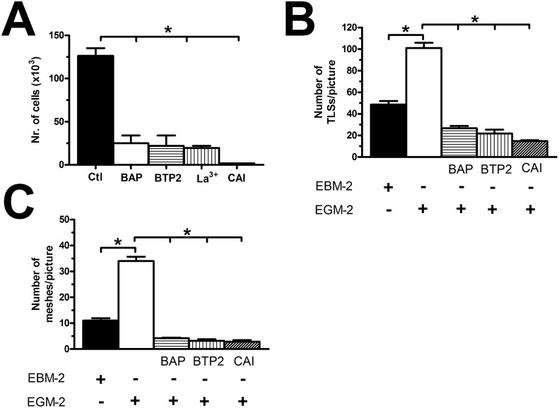
Figure 13. The pharmacological inhibition of store-operated Ca2+ entry blocks proliferation in breast cancer-derived endothelial colony forming cells.
(A), BAPTA (30 μM, 2 hours), a membrane-permeable buffer of intracellular Ca2+ levels, BTP2 (20 μM, 30 min), La3+ (10 μM, 30 min) and CAI (10 μM, 30 min) blocked proliferation in BC-ECFCs cultured in the presence of EGM-2. The asterisk indicates p<0.05. (B-C), statistical analysis of the dimensional (total TLSs per picture) and topological (total number of junctions between adjacent TLS and total number of meshes per picture) parameters of the capillary-like networks established by BC-ECFCs plated in Matrigel scaffolds in the presence and absence of BAPTA (30 μM, 2 hours), BTP2 (20 μM, 30 min), and CAI (10 μM, 30 min). In vitro angiogenesis was stimulated by plating the cells in the presence of EGM-2, while the EBM-2 medium (which is devoid of growth factors) was used as a control. The results are representative of three different experiments conducted on cells derived from three different donors. The asterisk indicates p<0.05.

