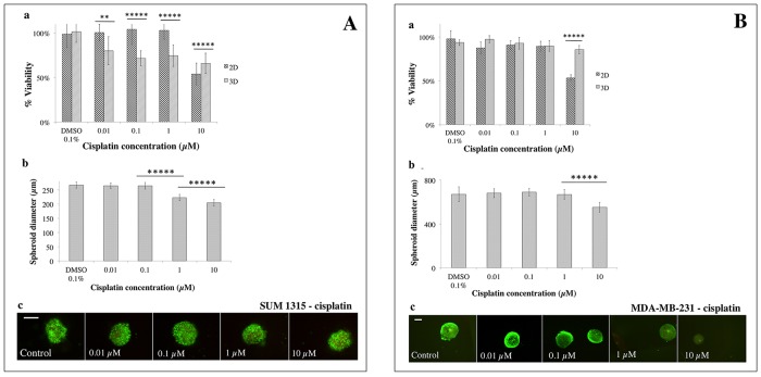
Figure 7. 2D vs 3D cell culture sensitivity to cisplatin.
(A) SUM1315 and (B) MDA-MB-231 cell line sensitivity to cisplatin (0.01, 0.1, 1, 10 μM) was assessed in 2D and 3D cell culture conditions after 5 days of treatment. (a) Cell viability in 2D and 3D culture conditions with the resazurin test: viability was calculated by the ratio of OD of resorufin formed in cisplatin-treated cells to 0.1% control DMSO cells. (b) Spheroid diameter measurement: this parameter was measured with ToupView® software (μm). (c) Live/Dead® spheroid imaging: Green marking corresponds to calcein-AM penetration (viable cells). Red markings correspond to ethidium homodimer-1 cell penetration (dead cells). Scale bar =200 μm. Results are displayed as mean ± SEM where *p<0.05, **p<0.01, ***p<0.001, ****p<0.0001 and *****p<0.00001.

