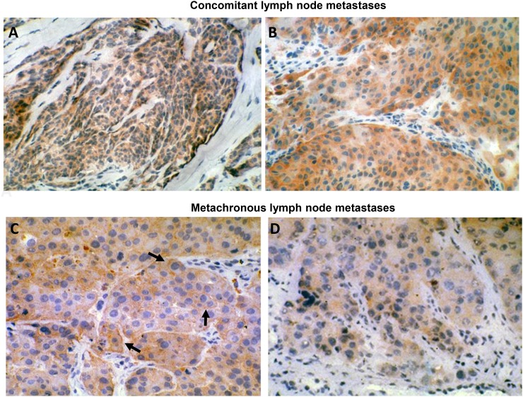
Figure 1. HER-3 expression in human metastatic melanoma.
(A) (160X original magnification) and (B) (250X original magnification): concomitant lymph node metastases. (C) (250X original magnification) and (D) (250X original magnification): metachronous lymph node metastases. Tissues were stained for HER-3 expression using MP-RM-1 antibody. Black arrows indicate MP-RM-1 membrane reactivity.

