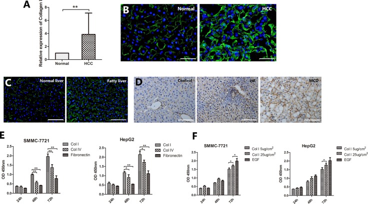
Figure 2. Collagen I contributes to the NAFLD-related HCC.
(A) qRT-PCR analysis showed that Collagen I levels were 2.8 fold higher in HCC samples than those in adjacent non-tumor liver tissues. (B) Immunofluorescence staining confirmed the higher expression of Collagen I (green) in HCC tissues compared to normal liver tissues. (C) Immunofluorescence staining revealed a higher expression of Collagen I (green) in human fatty liver compared to human normal liver. (D) In the mouse models of NAFLD/NASH, Collagen I was found to be upregulated. (E, F) HCC cells cultured on different ECM or different concentration of Collagen I to determine the effect of Collagen I on HCC cell proliferation. The vitality of cells at each time point was detected by CCK-8 assay. (E) SMMC-7721 and HepG2 cells cultured on Collagen I proliferated significantly faster than those on either Collagen IV or fibronectin. (F) The effect of Collagen I on cell proliferation appeared to be dose-dependent. Scale bar: 50 μm (B); 100 μm (C, D), *P < 0.05, **P < 0.001.

