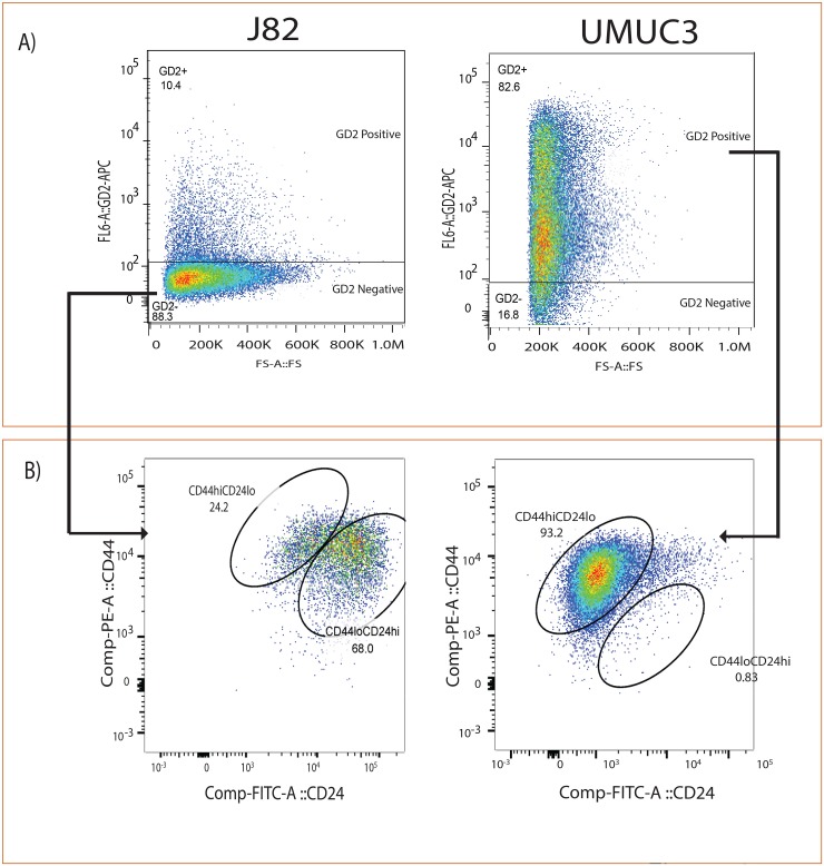
Figure 2. GD2 identifies CD44hiCD24lo in bladder cancer cells.
(A) Flow cytometry analysis of J82 and UMUC3 cells stained with anti-GD2-APC. (B) Anti-CD44-PE and anti-CD24-FITC antibodies cells were electrically gated on GD2+/– cells and displayed in a pseudo color dot plot with CD44 on the y-axis and CD24 on the x-axis using Flow Jo data analysis software.

