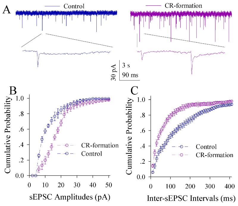
Figure 5. Excitatory synaptic transmission on the pyramidal neurons of the piriform cortices increases in CR-formation mice.
Spontaneous excitatory postsynaptic currents (sEPSC) were recorded on pyramidal neurons in cortical slices under voltage-clamp (holding potential at -70 mV) in the presence of 10 μM bicuculline. (A) illustrates sEPSCs recorded on a neuron from control mouse (blue trace in left panel) and CR-formation mouse (red in right). Bottom traces are the expanded waveforms selected from top traces. Calibration bars are 30 pA, 3 second (top) and 90 ms (bottom). (B) shows the cumulative probability of sEPSC amplitudes on the neurons from controls (blue symbols, n=15 neurons from nine mice) and CR-formation mice (red, n=15 neurons from nine mice). (C) illustrates the cumulative probability of inter-sEPSC intervals from controls (blue symbols, n=15 neurons from nine) and CR-formation mice (red, n=15 neurons from nine).

