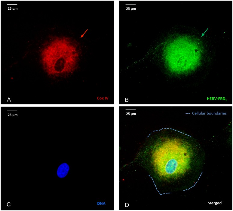
Figure 3. Stress-induced, co-localization of mitochondria and HERV-proteins.
Cellular localization of HERV proteins in U87RETO cells upon etoposide exposure at 5 μg/ml final concentration (for 10 days) measured by immunocytochemistry. The intracellular distribution of syncytin 2 (HERV- FRD1) was associated with mitochondria. Picture A: Cox IV staining, picture B: staining of HERV- protein using anti-syncytin 2-FITC conjugated antibody. Same results were obtained using anti-HERV-FITC conjugated antibodies against HERV-V3-1 and syncytin 1 (HERV-WE1), respectively (not shown). Picture C: negative control, DNA was labeled with DAPI, picture D: combined staining with all three merged channels. Magnification 40x. The figure is representative of at least n = 3 independent experiments.

