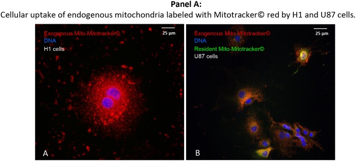
Figure 7. Direct, trans-membranous cellular uptake of purified and labeled mitochondria by cancer cells.
Picture A: the donor-mitochondria were previously isolated from U87RETO cells and labeled with red MitoTracker®. These prepared “red” mitochondria were added to H1RETO testicular carcinoma cells, whose endogenous mitochondria were not labeled. This effect was also detectable in wild-type U87 cells (data not shown). Picture B: the same prepared “red” mitochondria were added to U87RETO glioblastoma cells, whose endogenous mitochondria were previously labeled with green MitoTracker®. Incorporation was measured for at least 24 h. DNA was labeled with DAPI. Magnification 20x. The figure is representative of at least n = 3 independent experiments.

