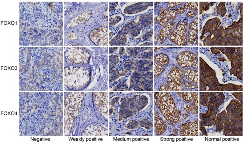
Figure 1. Expressions of FOXOs in cancer and paracancerous tissue, detected by immunohistochemistry.
The expressions of FOXO1, FOXO3 and FOXO4 are shown. The density of staining includes negative, weakly positive, moderately positive, strongly positive and normal positive. FOXO, forkhead box class O.

