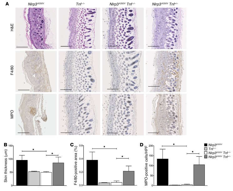
Figure 5. KO of TNF eliminates inflammatory skin disease in Nlrp3A350V mice.
(A–D) IHC of skin sections from Nlrp3A350V mice showed positive staining for F4/80 and MPO, with the presence of neutrophil pockets and a notable loss of s.c. tissue, while skin sections from Nlrp3A350V Tnf–/– mice had a complete absence of these and were indistinguishable from those of control animals. Nlrp3A350V Tnf+/– mice were partially protected, with less F4/80- and MPO-positive staining and increased s.c. tissue compared with skin tissue from Nlrp3A350V mice. Quantification of (B) skin thickness, (C) F4/80-positive cells, and (D) MPO-positive cells. Representative sections and quantification of skin thickness and IHC are from 9 mice in each group. Representative sections in A are oriented with the basal region on the far right of each panel (original magnification, ×10). Scale bar: 200 μm. *P < 0.05, by Kruskal-Wallis with Dunn’s multiple comparisons test. Data represent the mean ± SEM.

