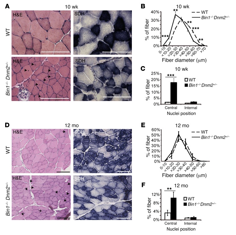
Figure 2. Skeletal muscle histology is mildly affected in surviving Bin1–/– mice with reduced DNM2 expression.
Transverse TA sections from 10-week-old (10 wk) (A) or 12-month-old (12 mo) (D) mice were stained with H&E or for SDH. Arrows point to mislocalized nuclei. Scale bars: 100 μm. H&E–stained muscle sections were then analyzed for fiber area for 10 wk (B) and 12 mo (E). Fiber diameter is grouped into 10-μm intervals, and represented as the percentage of total fibers. (C and F) The frequency of fibers with central or internal nuclei were scored. Internal nuclei are defined as neither subsarcolemmal nor central. All graphs depict the mean ± SEM. Statistical analysis was performed using an unpaired 2-tailed Student’s t test. **P < 0.01, ***P < 0.001, n = minimum 5 mice per group.

