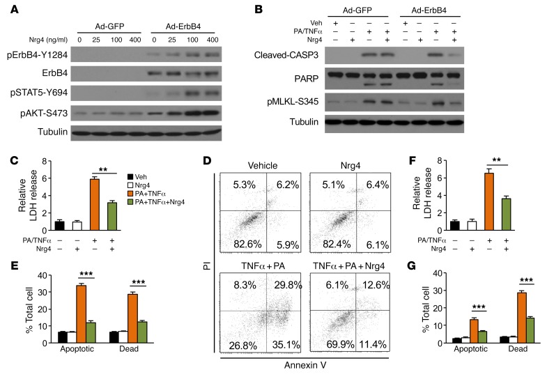
Figure 4. Nrg4 signaling protects hepatocytes from stress-induced cell death.
(A) Immunoblots of total lysates from primary hepatocytes transduced with GFP or ErbB4 adenovirus and treated with Nrg4 for 20 minutes. (B) Immunoblots of total lysates from primary hepatocytes transduced with GFP or ErbB4 adenovirus treated with 150 μM PA for 2 hours followed by addition of 40 ng/ml TNF-α (PA/TNF-α) and 100 ng/ml Nrg4 for 20 hours. (C) LDH activity in culture media from hepatocytes transduced with Ad-ErbB4 and treated as indicated for 20 hours. Data represent mean ± SEM. **P < 0.01, 1-way ANOVA. (D) Flow cytometry analysis of hepatocytes (20 hours treatment) following annexin V and PI staining. Double-positive cells are considered dead, whereas annexin V–positive and PI-negative cells are apoptotic. (E) Quantitation of hepatocyte cell death based on annexin V/PI staining. Data represent mean ± SEM. ***P < 0.001, 1-way ANOVA. (F) LDH activity in culture media from Hepa 1 cells stably expressing ErbB4 and treated as indicated for 20 hours. Data represent mean ± SEM. **P < 0.01, 1-way ANOVA. (G) Flow cytometry analysis of cell death in treated Hepa 1 cells stably expressing ErbB4 (20 hours treatment). Data represent mean ± SEM. ***P < 0.001, 1-way ANOVA.

