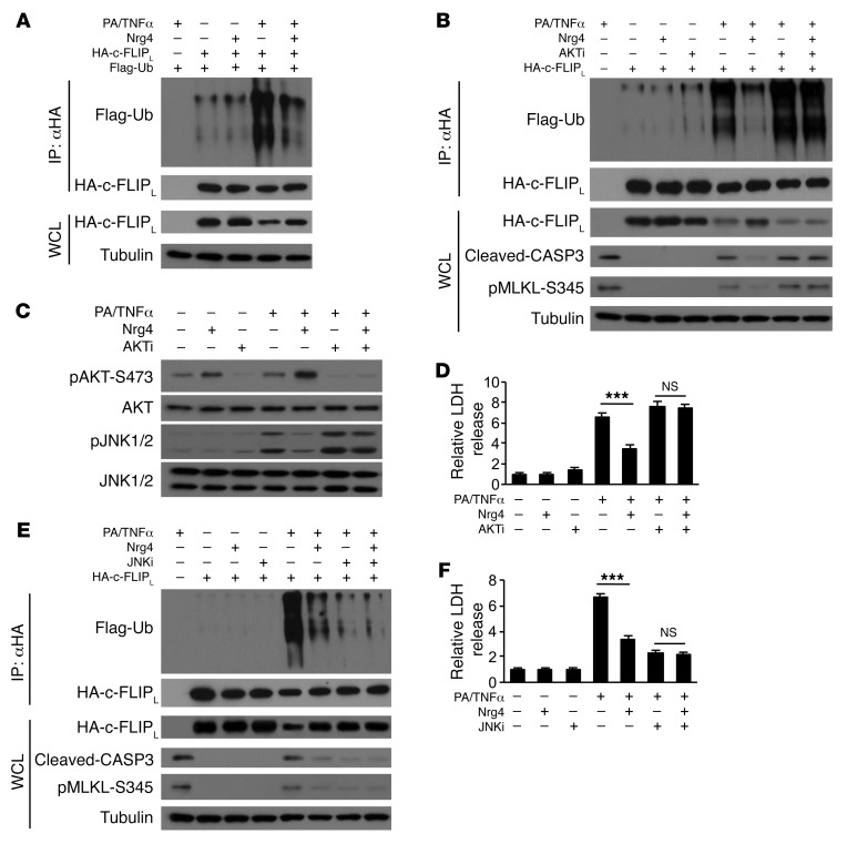
Figure 6. Nrg4 attenuates ubiquitination and proteasomal degradation of c-FLIPL.
The following experiments were performed in Hepa 1 cells stably expressing ErbB4. Cells were cultured in serum-free medium during treatment. WCL, whole cell lysates. (A) Immunoblots of IP and whole cell lysates from Hepa 1 cells transfected with plasmids encoding HA-tagged c-FLIPL and Flag-tagged ubiquitin (Flag-Ub) and treated with PA/TNF-α without or with 100 ng/ml Nrg4 for 4 hours. (B) Immunoblots of IP and whole cell lysates from transfected Hepa 1 cells treated in the absence or presence of AKT kinase inhibitor (20 μM) for 4 hours. (C) Immunoblots of total lysates from cells treated with PA/TNF-α without or with 100 ng/ml Nrg4 and in the absence or presence of AKT inhibitor (20 μM) for 6 hours. (D) LDH release by Hepa 1 cells as treated for 20 hours. Data represent mean ± SEM. ***P < 0.001, 1-way ANOVA. (E) Immunoblots of IP and WCL from transfected Hepa 1 cells treated in the absence or presence of JNK1/2 kinase inhibitor (20 μM) for 4 hours. (F) LDH release by Hepa 1 cells as treated for 20 hours. Data represent mean ± SEM. ***P < 0.001, 1-way ANOVA.

