
Fig. 1.
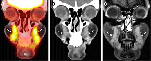
45-year-old female patient evaluated with PET-CT for lymphoma staging. Coronal PET-CT image (a) reveals incidentally detected FDG avid areas in bilateral nasolabial fat compartments (arrows). SUVmean = 4.6, SUVmax = 5.9. The areas appear mildly hyperdense on CT (arrows in b) and hardly enhancing on post-gadolinium T1 W fat saturated sequences (c, arrows). The patient had a history of silicone injections four years earlier. She had no filler-related symptoms