
Fig. 8.
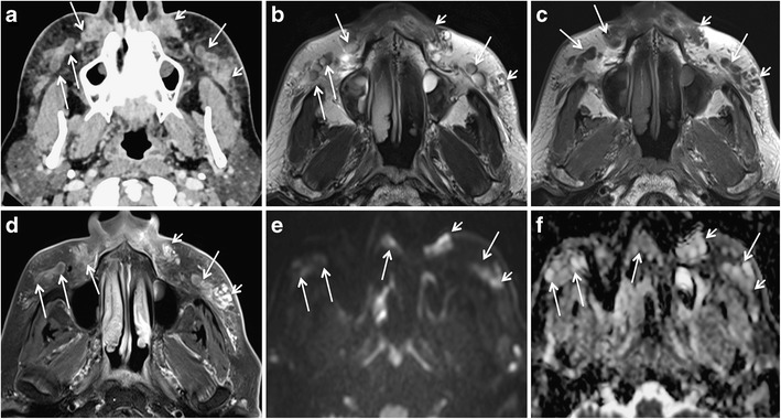
A 65-year-old woman developed pain, erythema, and bilateral cheek swelling after 6 months of HA facial filler injections. a Contrast-enhanced CT shows bilateral “grape-like” hypodense, rim-enhancing areas (long arrows) and solid appearing enhancing nodules (short arrows) in the nasolabial fat, medial and middle superficial cheek fat compartments. T2 W (b), T1 W (c), and fat-saturated gadolinium enhanced T1 W (d) images reveal that the rim-enhancing areas already seen on CT have a high protein content (hypo-isointense on T2 and T1). On b1000 (e) and ADC map (f), these rim-enhancing lesions show variable diffusivity (long arrows). ADC values were between 1.2 and 2 × 10⁻³ mm2/s. The solid lesions (short arrows) show strong enhancement on MRI and no restricted diffusivity. Surgery confirmed bilateral infected fluid collections and isolated FBG