Fig. 9.

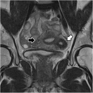
Isolated or juvenile cystic adenomyoma: Coronal T2-weighted images; nodular uterine lesion with a central cavity with hyperintense signal (white arrow), without connection to the endometrial cavity in an otherwise normal uterus (black arrow)

Isolated or juvenile cystic adenomyoma: Coronal T2-weighted images; nodular uterine lesion with a central cavity with hyperintense signal (white arrow), without connection to the endometrial cavity in an otherwise normal uterus (black arrow)