
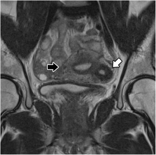
Fig. 9.
Isolated or juvenile cystic adenomyoma: Coronal T2-weighted images; nodular uterine lesion with a central cavity with hyperintense signal (white arrow), without connection to the endometrial cavity in an otherwise normal uterus (black arrow)
Official websites use .gov
A
.gov website belongs to an official
government organization in the United States.
Secure .gov websites use HTTPS
A lock (
) or https:// means you've safely
connected to the .gov website. Share sensitive
information only on official, secure websites.
Isolated or juvenile cystic adenomyoma: Coronal T2-weighted images; nodular uterine lesion with a central cavity with hyperintense signal (white arrow), without connection to the endometrial cavity in an otherwise normal uterus (black arrow)