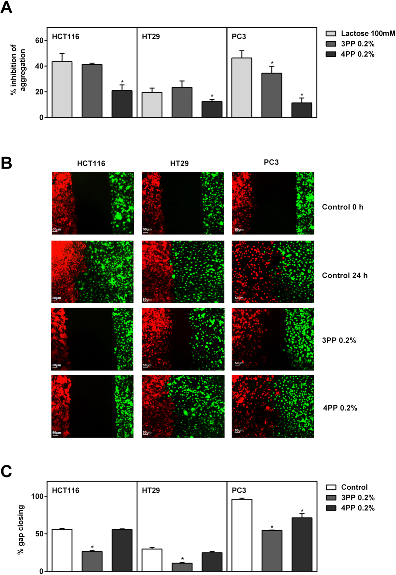
Figure 3.
Homotypic aggregation and migration assays (wound healing and endothelial vs cancer cells). (A) Inhibition of homotypic cell aggregation using asialofetuin treated with lactose or papaya pectin at 0.2%. 3PP strongly inhibited cancer cells aggregation. The results were expressed in percentage of cells in relation to control (with asialofetuin and no treatment). Data were shown as mean ± SD from two independent WSF samples, each one performed in technical duplicate, from the biological duplicate (n = 4). *P < 0.05 vs lactose, according to Dunnett’s test. Images of homotypic aggregation test were in Supplementary Figure S1. (B) Endothelial cells (BAMEC) dyed with DiO (green) and cancer cells dyed with DiI (red). 3PP diminish the interaction between cancer cells and BAMEC. Scale bar: 50 µm. Representative image of, at least, two experiments from the biological samples. (C) Quatification of gap closing after 24 h. 3PP slowest gap closing compared with control and with 4PP. The results were expressed in percentage of cells that invaded the gap compared with control. Data were shown as mean ± SD (as previously explained in Figure 3A). *P < 0.0001 vs control (without treatment), according to Dunnett’s test. Images of wound healing were in Supplementary Figure S2. PP: papaya pectin (water-soluble fraction).

