Figure 1.

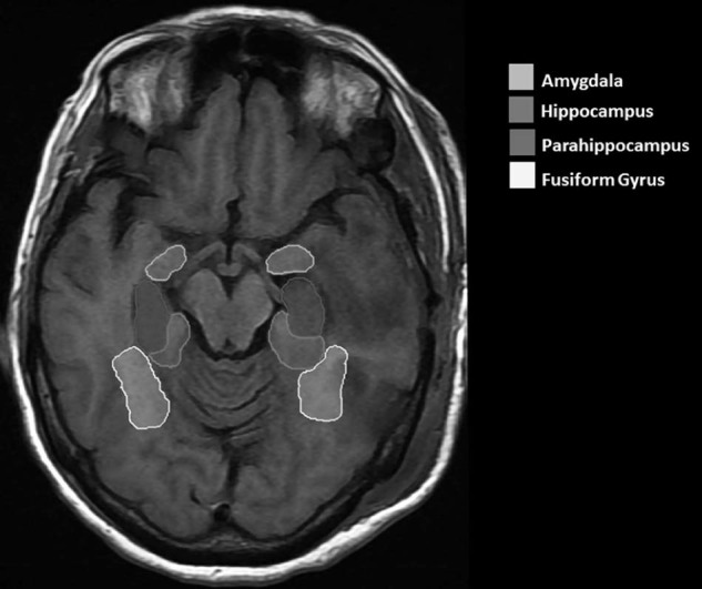
Delineation of regions of interest on T1-weighted magnetic resonance imaging of the brain. The hippocampus, amygdala, parahippocampus, and fusiform gyrus are delineated. All structure volumes included both the left and right sides.

Delineation of regions of interest on T1-weighted magnetic resonance imaging of the brain. The hippocampus, amygdala, parahippocampus, and fusiform gyrus are delineated. All structure volumes included both the left and right sides.