Fig. 4.
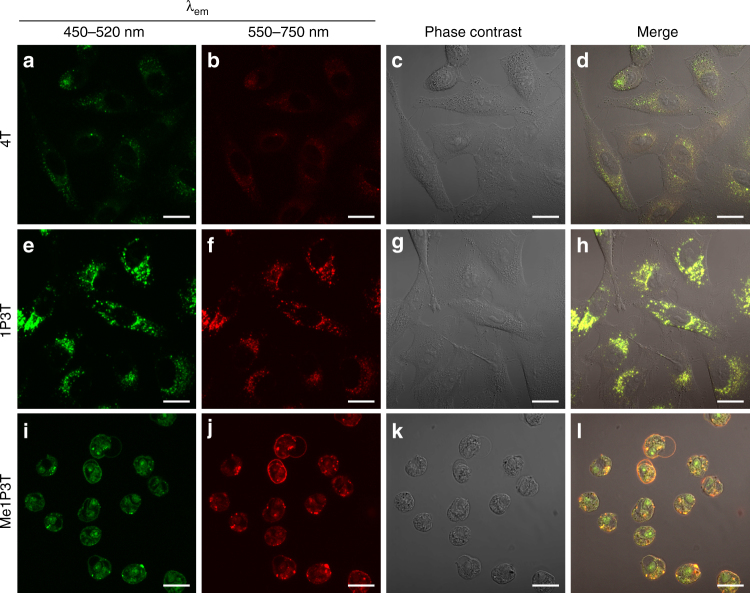
Co-localized cellular uptake and morphologies of A549 cells with free oligothiophenes. Confocal laser scanning micrographs of A549 cells treated with 15 µM oligothiophenes 4T a–d, 1P3T e–h, and Me1P3T i–l for 24 h at 37 °C, 5% CO2. All samples are excited at 405 nm, where the molecular states (450–520 nm, green) and the aggregated states (550–750 nm, red) of the oligothiophenes were visualized simultaneously. Co-existence of both aggregated and non-aggregated species was represented as yellow signals. Scale bar = 20 µm. Poor cellular internalization was observed for 4T d, whereas co-localization of molecular and aggregated species were detected for 1P3T h. Significant cell rounding indicating cellular toxicity was observed for Me1P3T l

