Figure 6.
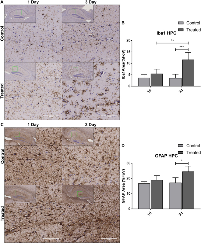
Topical Aldara application induces significant changes in Iba1+ and GFAP+ cells in the hippocampus. Immunohistochemistry was performed on formalin-fixed paraffin embedded brain sections from mice treated daily with Aldara or control cream. Samples were collected 24 hr after first treatment and 24 hr after 3 daily treatment. (A) Representative images of Iba1+ staining in mouse hippocampus. (B) Quantification of Iba1+ area in mouse hippocampus. (C) Representative images of GFAP+ staining in mouse hippocampus. (D) Quantification of GFAP+ area in mouse hippocampus. Quantification was performed using ImageJ analysis software. Significance was determined using one-way ANOVA with post-hoc Student’s t-test with Bonferroni’s multiple testing correction. *P < 0.05; **P < 0.01; ***P < 0.001.

