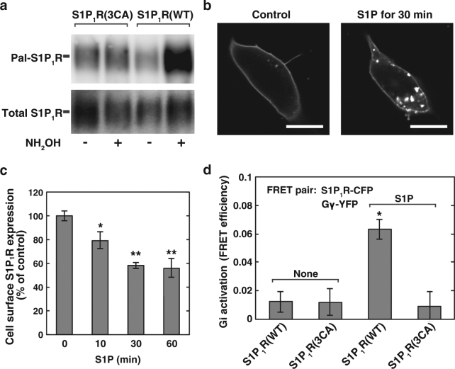
Figure 5.
Palmitoylation-deficient mutant S1P1R(3CA) was unable to couple with Gi. (a) SH-SY5Y cells transiently expressing wild-type S1P1R-FLAG or S1P1R(3CA)-FLAG were analysed for palmitoylation by ABE assay. One of the representative results from three independent experiments is shown. Full-length images are presented in Supplementary Figure 9. (b) SH-SY5Y cells transiently expressing S1P1R(3CA)-CFP were stimulated with or without 100 nM S1P for 30 min and fixed with paraformaldehyde. The localisation of S1P1R(3CA)-CFP was analysed by confocal microscopy. Bars, 10 µm. One of the representative results from three independent experiments is shown. (c) SH-SY5Y cells transiently expressing S1P1R(3CA)-CFP were stimulated with 100 nM S1P for various time periods as specified. After fixation of the cells, the fluorescence of each protein was analysed by confocal microscopy. Fluorescence intensity at each time point was quantitated and compared with time point 0 (control). Values represent means ± s.e.m. of 3 independent experiments carried out in triplicate. Statistical significance was analysed by Student’s t-test (n = 9; **P < 0.01, *P < 0.05 versus control (0 min)). (d) SH-SY5Y cells transiently expressing wild-type S1P1R-CFP or S1P1R(3CA)-CFP together with Gβ and Gγ-YFP were stimulated with or without 100 nM S1P for 2 min, fixed and analysed for FRET efficiencies. Values represent means ± s.e.m. (n ≥ 50). Statistical significance was analysed by Student’s t-test (*P < 0.05 versus wild-type S1P1R with S1P).

