. 2015 Apr 8;6(7):3839–3844. doi: 10.1039/c4sc03894k

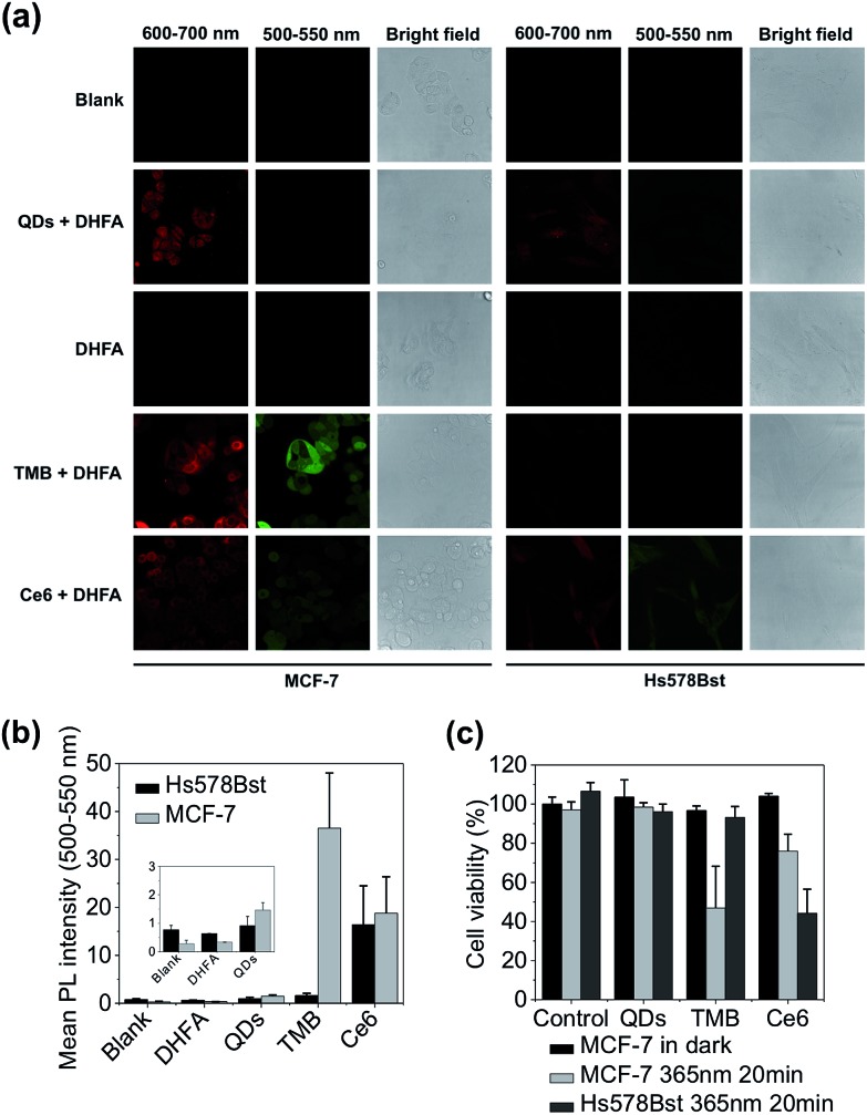
Fig. 4. Evaluation of theranostic efficacy of TMB with live cancer cells. (a) Confocal microscopy images of MCF-7 and Hs578Bst cells incubated with QDs, TMB, and Ce6 and further treated with DHFA for intracellular 1O2 detection. Fluorescence signals (red) of each probe were collected between 600 nm and 700 nm with 405 nm excitation. DHFA fluorescence (green) was collected between 500 nm and 550 nm with 488 nm excitation. (b) Mean DHLA PL intensities of MCF-7 and Hs578Bst cells treated with QDs, TMB, and Ce6. (c) Cell viabilities of MCF-7 and Hs578Bst cells incubated with QDs, TMB, and Ce6. Cells were incubated with each probe for 3 hours. PDT was implemented with 365 nm UV light irradiation for 20 min.

Fig. 4