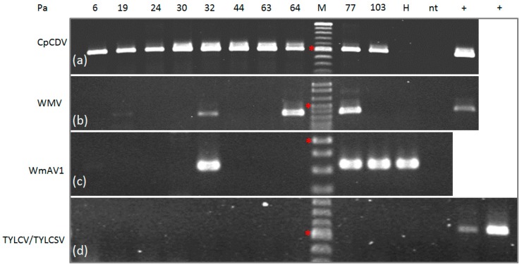
Figure 3.
Validation of virus infection in individual watermelon fruit sample used for NGS analysis; polymerase chain reaction (PCR) (a,d) and reverse transcription PCR (RT-PCR) (b,c); amplified fragments were separated by electrophoresis in 1.5% agarose gels in 0.5× TBE: (a) CpCDV, 501 bp amplified fragment; (b) WMV, 379 bp amplified fragment; (c) WmAV1, putative novel amalgavirus, 333 bp amplified fragment (no positive control is available); (d) TYLCV/TYLCSV, 580 bp amplified fragment, positive controls: TYLCV (+left) and TYLCSV (+right). H, seed-borne watermelon fruit; nt, no template; +, positive controls; M, 100 bp DNA Ladder, (*) indicates the 500 bp fragment.

