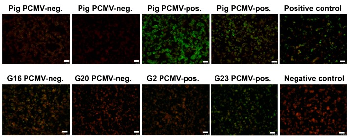
Figure 5.
Results of the immunofluorescence analysis of pig and human sera for antibodies reacting against HHV-6. HHV-6 infected cells were used as antigen. In the upper row the reactivity of four pig sera is shown, two of them were PCMV-negative two were PCMV-positive. The “positive control” and the “negative control” are human sera with a known seroreactivity. In the upper row four human sera were analyzed which in parallel were tested on a Western blot analysis using four recombinant proteins of PCMV (Table 5). The bar corresponds to 50 µm.

