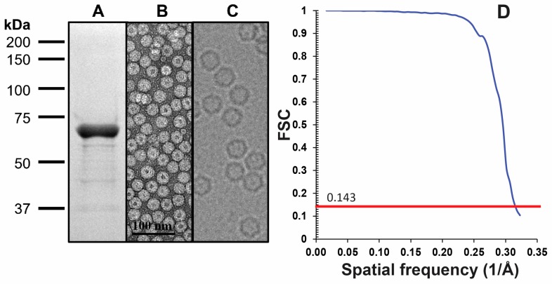
Figure 1.
LuIII sample and data quality. (A) SDS-PAGE of purified VLPs. The VP2 is ~65 kDa in size; (B) negative stain EM of purified VLPs (42,000×, Scale bar = 100 nm); (C) representative micrograph from cryo-EM data collection, not visualized to scale; (D) FSC plot for the final iteration of 3D map reconstruction (red line indicates the 0.143 threshold utilized to estimate resolution).

