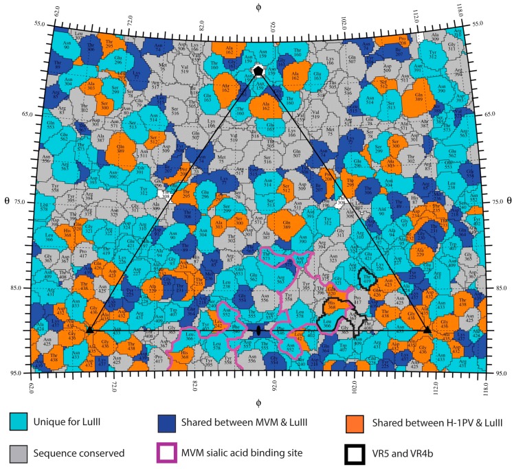
Figure 6.
Distribution of amino acid sequence identity for the rodent protoparvoviruses. 2D projection roadmap of the LuIII capsid surface showing the viral asymmetric unit (black triangle). Colors are as indicated.VP2 positions containing an amino acid that is unique to LuIII are in cyan. Residues identical between: MVM and LuIII (blue), H-1PV and LuIII (orange), or conserved among all three viruses (gray). The remaining residues in white are identical between H-1PV and MVM only. The sialic acid binding site identified for MVM and predicted for H-1PV is delineated in purple (see bold outline). Important surface exposed residues within VR5 and VR4b are delineated in black. Icosahedral axes are indicated by the filled oval (2-fold), triangle (3-fold), and pentagon (5-fold).

