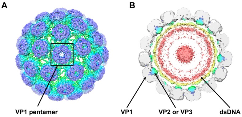
Figure 1.
Cryo-electron microscopy structure of BK virus (BKPyV) viral particles (Adapted from [11]). (A) External view of the BKPyV virion shown at a contour level of 0.022. A viral protein VP1 pentamer is highlighted; (B) View of a 40-Å-thick slab through the unsharpened/unmasked virion map shown at a contour level of 0.0034. Pyramidal density below each VP1 penton and two shells of electron density adjacent to the inner capsid layer can be seen. The density within 6 Å of the fitted coordinates for SV40 VP1 is coloured grey. Density for VP2 and VP3 is coloured blue/green and for packaged double stranded DNA (dsDNA) yellow/pink.

