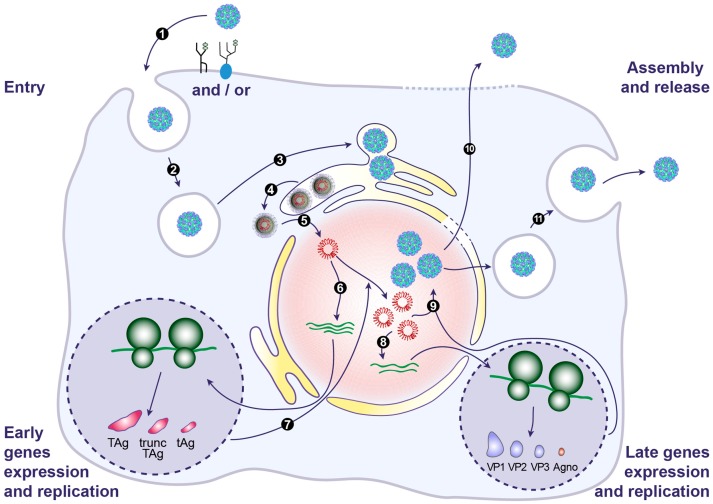
Figure 3.
Model of the BKPyV life cycle. BKPyV infection begins with binding of virions to the ganglioside receptors (particularly GT1b and GD1b) and/or an N-linked glycoprotein containing α(2,3)-linked sialic acid, at the cell surface (1). This is followed by internalization potentially through a caveola-mediated endocytosis step within the first 4 h after adsorption (2). The virus subsequently traffics from the late endosomes to the endoplasmic reticulum (ER), where it arrives approximately 10 h post-infection (3). In the ER, virions benefit from chaperones, disulphide isomerases and reductases to facilitate the partial capsid uncoating. This creates a hydrophobic surface exposing VP2/VP3 that binds to and integrates into the ER membrane, leading to the release of partially uncoated viruses into the cytosol, a process that also involves the ER-associated protein degradation (ERAD) machinery (4). The viral genome is then transported into the nucleus via the nuclear pore complex thanks to VP2/VP3 NLS and the importin α/β1 import pathway (5). Expression of early genes occurs approximately 24 h post-infection (6). Early proteins are translocated into the nucleus where they serve to initiate viral DNA replication (7). Late genes are then expressed (8). VP1, VP2 and VP3 are translocated into the nucleus where they self-assemble to form capsids into which newly synthetized double stranded viral DNA is packaged (9). Progeny virions are mainly released from infected cells after cell lysis (10). However, a small fraction of progeny virions may also be released into the extracellular environment through a non-lytic egress that depends on the cellular secretion pathway (11).

