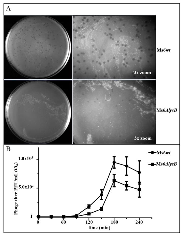
Figure 2.
(A) Phage plaques formed by Ms6 (top) or Ms6ΔlysB (bottom) on a lawn of M. smegmatis. The plaques formed by Ms6ΔlysB phage are smaller than the ones formed by the wild-type Ms6; (B) one-step growth curves of Ms6wt (circles) or Ms6ΔlysB (squares) on M. smegmatis mc2155 show a lower number of plaque-forming units (PFU) released from Ms6ΔlysB infection. Both curves show similar progression up to 90 min post-adsorption showing no differences in the timing of lysis. T0 marks the end of the adsorption and start of the one-step experiment. The PFU/mL at t = 0 was used to normalize PFU/mL of each time point. For each time point, the mean ± SD of four independent assays is indicated.

