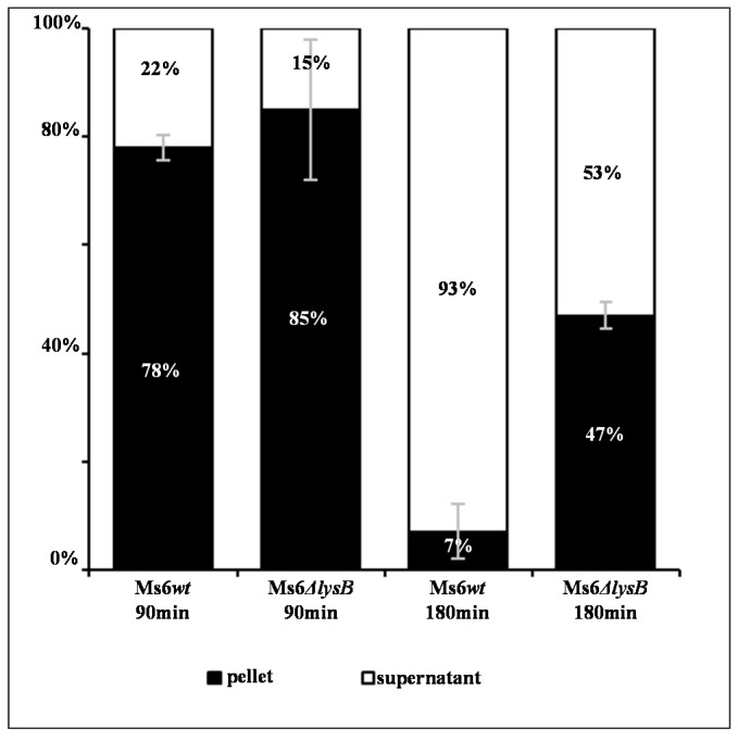
Figure 3.
Distribution of phage particles in the supernatant and pellet of M. smegmatis infected with Ms6wt or Ms6ΔlysB. Ms6 is trapped in cell debris in absence of LysB. At the indicated time points, the distribution of phage particles in the pellet and in the supernatant was determined as a percentage of the total amount of PFU counted in both fractions. The values indicate the mean ± SD of three independent experiments.

