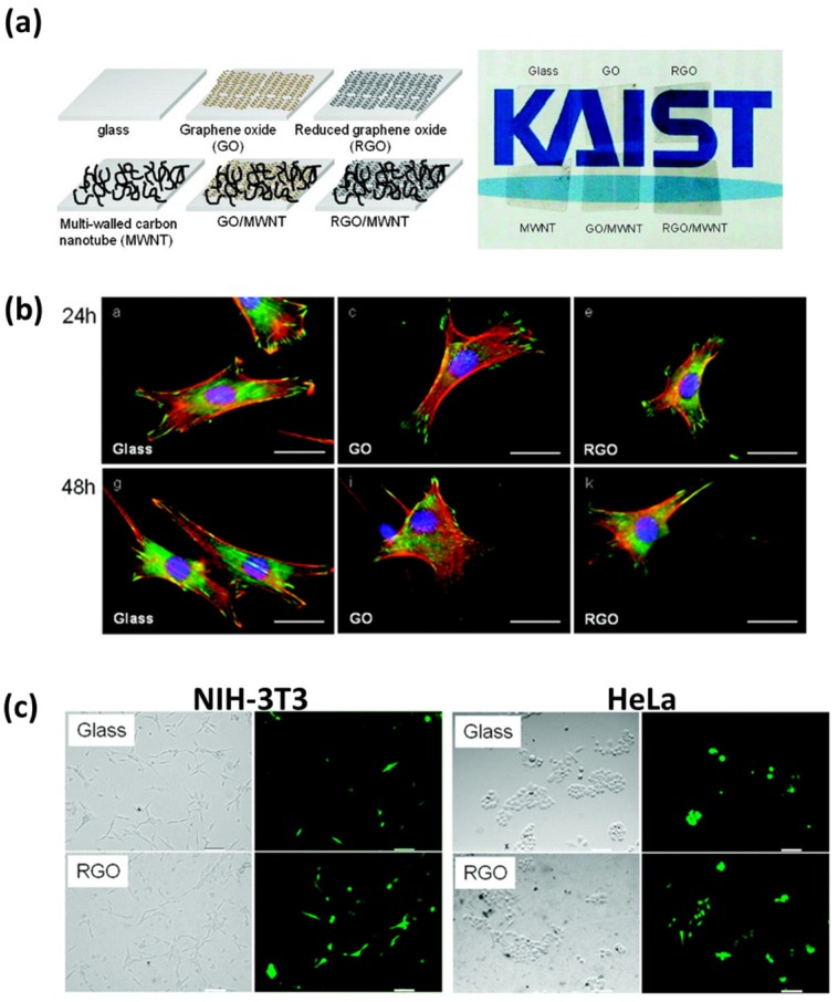
Figure 1.
Stimulating effects of graphene nanomaterial-coated substrates on NIH-3T3 fibroblast behaviors. (a) Structural diagrams (left panel) and optical images (right panel) of graphene nanomaterial-coated substrate. (b) The fluorescence images of NIH-3T3 fibroblasts on each substrate for 24 and 48 h. Scale bars are 20 μm. (c) Improved gene transfection efficiency of NIH-3T3 fibroblasts and HeLa cells on rGO-coated substrates after 48 h incubation. Scale bars are 35 μm. Reproduced with permission from [44]. American Chemical Society, 2010.

