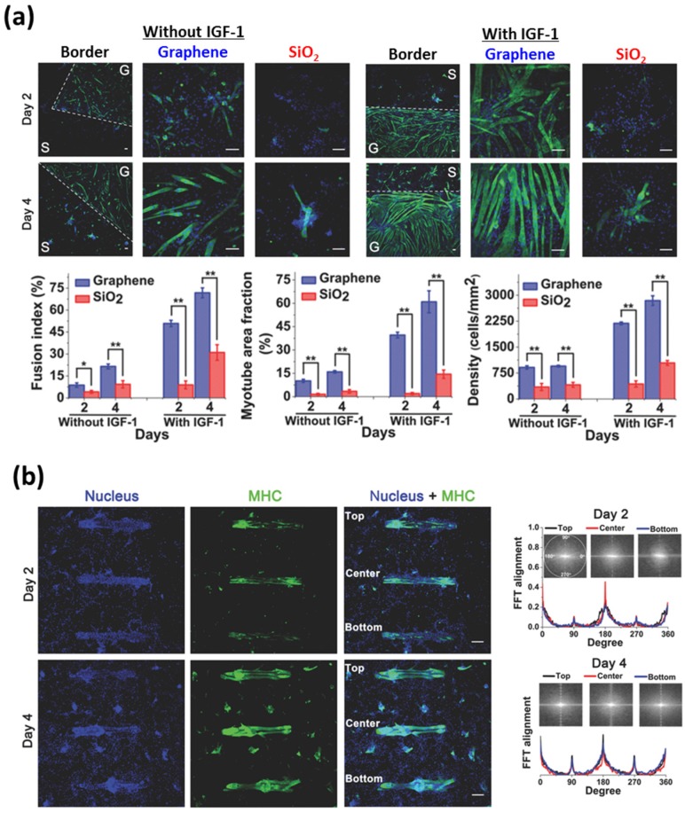
Figure 5.
Graphene-based patterning and differentiation of myoblasts. (a) Myogenic differentiation of C2C12 skeletal muscle myoblasts on graphene-patterned substrates. The dashed white line in column one indicates the border of SiO2 and graphene surfaces on the substrates. Scale bars are 100 μm. (b) Fluorescence images and alignment of the C2C12 myotubes on rectangular island-shaped graphene patterns. Scale bars are 250 μm. Reproduced with permission from [71]. Copyright John Wiley and Sons, 2013.

