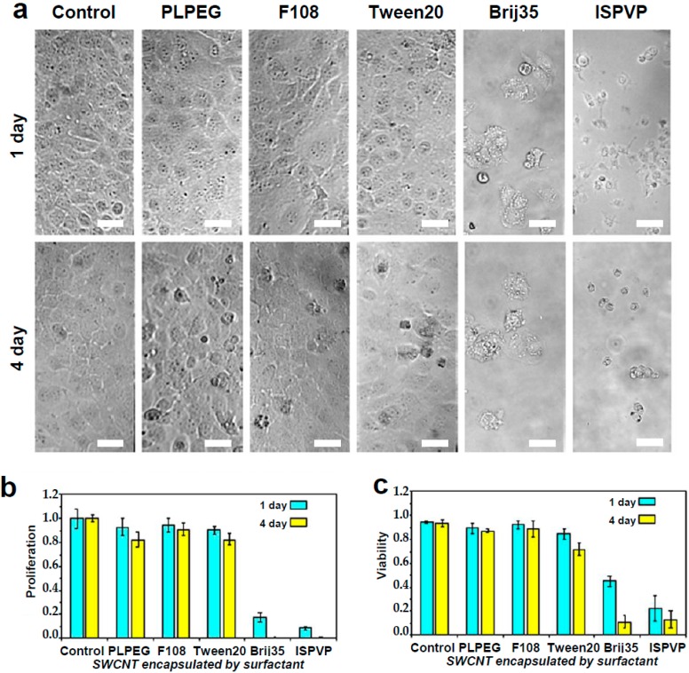
Figure 1.
Live cell biocompatibility of SWCNTs encapsulated with different coatings. Top (a): Bright-field images of COS-7 incubated with PLPEG-, F108-, Tween20-, Brij35-, and ISPVP-coated SWCNTs for one day and four days. Scale bar: 30 µm. Bottom: Corresponding comparisons of cellular (b) proliferation and (c) viability. Starting concentration of COS-7 cells: 1 × 10⁵ cells/mL, SWCNT: 1 μg/mL, cell cultured at 37 °C with 5% CO2; Three independent experiments were performed to obtain standard variations. Cell viability was evaluated using trypan blue dye staining.

