Figure 2.
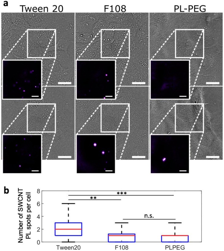
SWCNT interactions with live cells probed by NIR photoluminescence. (a) Bright-field and NIR photoluminescence imaging (inserts) of live cells incubated for 24 h with Tween20-, F108-, or PLPEG-coated SWCNTs and further rinsed before imaging. PLPEG- and F108-coated SWCNTs displayed lower non-specific interactions with live cells compared to Tween20-coated SWCNTs. Scale bars: 25 µm for the bright field images and 10 µm for the magnified NIR photoluminescence images of SWCNTs; (b) Corresponding median (red), 25–75th percentile (blue), and 0–100th percentile (black) of the number of SWCNT PL spots observed on live cells for Tween20-, F108- or PLPEG-coated SWCNTs (N = 70, 53, and 86 cells respectively, n.s.: not significant, ** p < 0.01, *** p < 0.001, Kolmogorov-Smirnov test).

