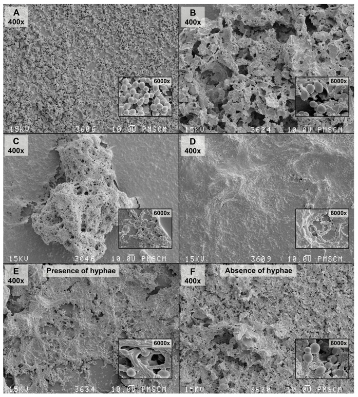
Figure 8.
SEM images of the mono-species biofilm generated by C. albicans, S. mutans and the double-species oral streptococci/yeasts biofilm—untreated and treated with the Lactobacillus salivarius (HM6 Paradens)—after 24 h of biofilm formation. (A) The 24 h C. albicans biofilm formed on a flat agar surface (Agar Scientific, Stansted, UK); (B) The 24 h C. albicans biofilm formed on a flat agar surface treated with the Lactobacillus salivarius (HM6 Paradens); (C) The 24 h S. mutans biofilm formed on a flat agar surface (Agar Scientific, UK); (D) The 24 h S. mutans biofilm formed on a flat agar surface treated with the Lactobacillus salivarius (HM6 Paradens); (E) Co-culture oral streptococci/yeasts biofilm—untreated with the Lactobacillus salivarius (HM6 Paradens)—after 24 h of biofilm formation; (F) Co-culture oral streptococci/yeasts biofilm—treated with the Lactobacillus salivarius (HM6 Paradens)—after 24 h of biofilm formation. The Culture was maintained at 36 °C, pH 7.0 and pCO2 5%, in bovine serum as a medium additive promoting growth of the culture in the presence of a sucrose substrate (5%). The occurrence of a co-culture biofilm at this stage may depend on the C. albicans morphotypes showing a twofold nature: buds and C. albicans hyphae may colonize mucous membranes and constitute physiological microflora (commensal) or may lead to infection under favorable conditions (opportunistic pathogens). There was no clear, compact structure for the C. albicans biofilm and single loosely located budding cells. Other pathological forms of yeasts are invisible. There was an apparent change in the C. albicans morphotype in the S. mutans common culture and visible pleomorphic forms were true hyphae, blastoconidia, which in the mixed culture also produce mycelial forms, whose role is related to damage to immune cells (macrophages) leading to microorganism invasion. There was abundant extracellular matrix between cells and covering bacterial and yeast cells. Bacterial cells were visible in chains adhering to yeast cells and wrapped around them. There was no clear, compact S. mutans/C. albicans biofilm structure or single loosely located budding cells. Other morphological forms of C. albicans were invisible. There was no visible extracellular matrix (D,F), which is formed by S. mutans alone and S. mutans with C. albicans treated with the Lactobacillus salivarius (C,E). Original magnification: 400×, 4000× and 6000×.

