Figure 2.
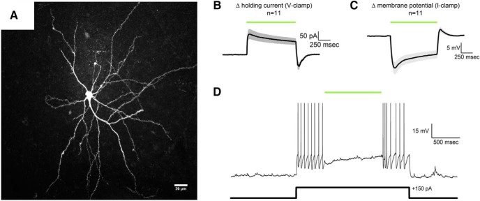
Functional validation of eNpHR3.0 in the BLA. A, Two-photon z-series projection of a mCherry-positive BLA neuron filled with biocytin and immunolabeled with Alexa-594. B, Activation of eNpHR3.0 produced a clear outward current in mCherry-positive BLA neurons (n = 11) voltage-clamped at −70 mV. Green bar represents optical stimulation. Shaded area around the average trace represents the SEM. C, Identical activation of eNpHR3.0 in current clamp produced robust hyperpolarization (n = 11). A brief, mild, rebound depolarization was apparent immediately after optical stimulation. This current, likely mediated by HCN channels (Womble and Moises, 1993; Park et al., 2007, 2011; Giesbrecht et al., 2010), had a mean amplitude in current clamp of 4.7 ± 1.3 mV and was almost always insufficient to drive the cells to threshold for action potentials. Green bar represents optical stimulation. Shaded area around the average trace represents the SEM. A small subset of individual sweeps that did have at least one rebound action potential after sustained eNpHR3.0-mediated hyperpolarization were removed from the average traces presented in B, C. D, A representative mCherry-positive BLA neuron that was current-clamped at 0 pA shows an increase in firing rate upon injection of a 150 pA current pulse, which is effectively suppressed during activation of eNpHR3.0.

