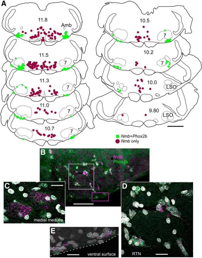
Figure 11.
In rats Nmb transcripts are present both in RTN and in the adjacent medial reticular formation. A, Drawing of coronal sections through medulla and caudal pons of rat showing the distribution of neurons with Nmb transcripts, with or without Phox2b transcripts. As in the mouse, Nmb + Phox2b neurons delineate the RTN. However, a large separate population of Nmb neurons is located more medially in rat. Numbers in center of sections indicate mm behind bregma (after Paxinos and Watson, 2005). Abbreviations as in Fig. 1. B, Photomicrograph of coronal section in caudal RTN showing transcripts for Nmb in magenta and Phox2b in green. DAPI stain is white/gray. Medial to the left, dorsal toward the top. C, Photomicrograph from midline region where Phox2b is not expressed in Nmb neurons. D, Enlargement of white dashed box from B. Note coexpression of Nmb with Phox2b transcripts. E, Enlargement of magenta dashed box in B showing cells expressing both Phox2b and Nmb transcripts. Scale: A, 500 μm. Scale bars: A, 1 mm; B, 100 μm; C–E, 20 μm.

