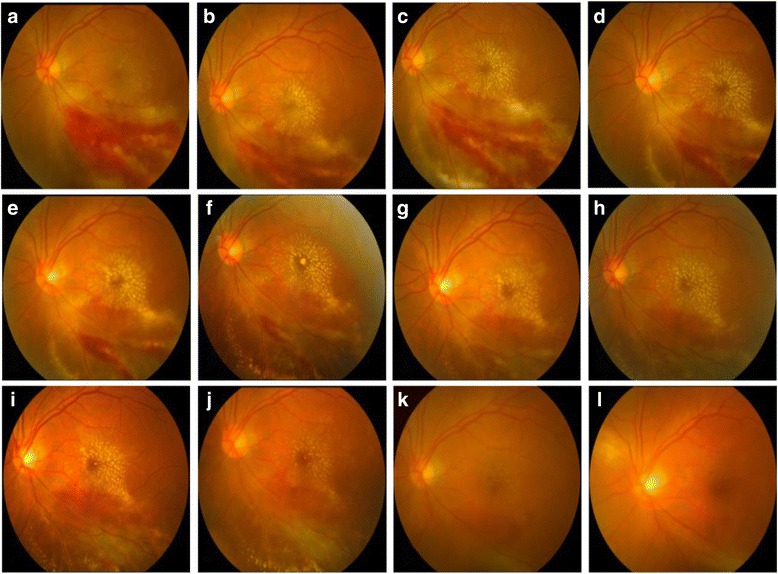
Fig. 1.

Fundus photography of the left eye showing the complete resolution of active retinitis after intravitreal injection of ganciclovir. a Left eye fundus photography showing active cytomegalovirus retinitis lesions at presentation. There is macular involvement. Initial visual acuity was 0.3 LogMAR. b Fundus photograph at the 3rd day after the first intravitreal ganciclovir injection in the left eye. c Fundus photograph at the 7th day after the second intravitreal ganciclovir injection in the left eye. d-j Fundus photography showing the gradually resolution of active lesions, with the remission of retinitis after treatment of weekly intravitreal ganciclovir injection. k Fundus photographs showing total resolution of active lesions at the 2nd month after the last intravitreal ganciclovir injection. l The fundus photography at the 5th month follow-up. Final visual acuity was 0.1 LogMAR
