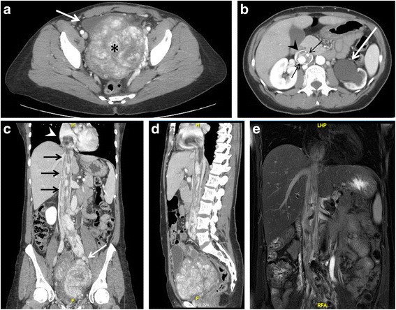
Fig. 3.

Chest – abdomen – pelvis CT scan (a-d) and MR (e). a. Axial projection showing a voluminous pelvic mass of 15 × 22 × 9 cm (asterisk), with significant contrast enhancement due to hypervascularization, causing right antero-lateral dislocation of the bladder (arrow). b. The mass also determines left hydro-ureteronephrosis (white arrow). Notice the presence of contrast-enhanced large vessels (black arrows) within the IVC (arrowhead). c-d. Coronal and sagittal CT projections showing the pelvic tumor directly extending into the left iliac vein (white arrow), IVC (black arrows) and occupying the right atrium (arrowhead). e. Coronal MR projection showing the caval and cardiac extension of the tumor
