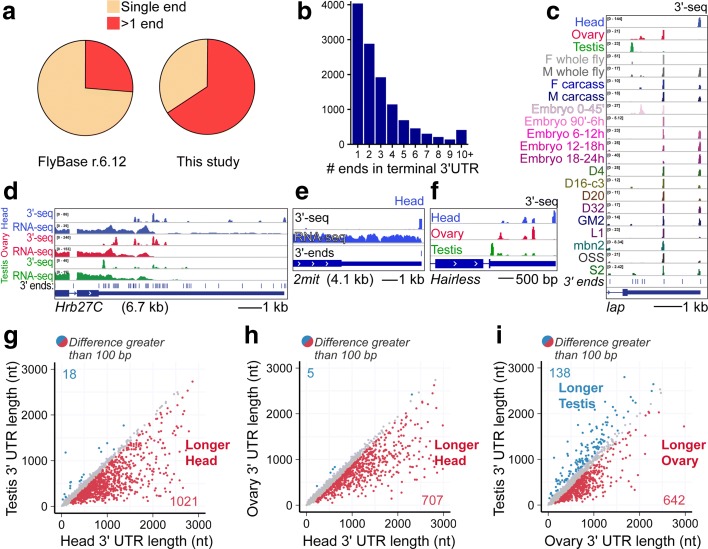
Fig. 2.
Tissue-specific alternative polyadenylation in D. melanogaster. a Fraction of genes with one or more annotated 3′ ends in the current FlyBase annotation (r6.12) compared to our revised 3′-seq-based atlas. b Summary of 3′ terminal isoform numbers annotated for genes in our 3′-seq-based atlas. c–f Examples of genes with diverse APA patterns. c Example of a gene with complex pattern of 3′ end isoform expression across adult tissues, embryonic timecourse, and cell lines. d Example of a gene with a long 3′ UTR that exhibits extreme 3′ end diversity. e Example of a gene with a long (4 kb) 3′ UTR that generates a single 3′ end in head. f Example of a gene with distinctive tissue-specific APA isoforms deployed in testis, ovary, and head. g–i Genome-wide patterns of tissue-specific APA revealed by pairwise analyses of weighted 3′ UTR length (denoted as 3′ UTR length for simplicity) comparison between tissues. Weighted 3′ UTR length is obtained taking the average of all 3′ UTR isoform lengths per gene weighted by the contribution of each isoform expressed. Genes are expressed at a minimum of 5 RPM in all samples. The genes for which weighted 3′ UTR length differs by 100 bp or more between samples are shown colored: red, longer weighted length in the sample on the x-axis; blue, longer weighted length in the sample on the y-axis. g Female head vs. testis. h Female head vs. ovary. i Ovary vs. testis

