Figure 2.

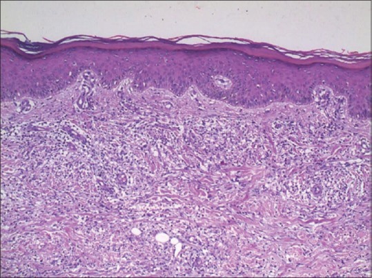
Photomicrograph showing lymphohistiocytic infiltrate extending in between the collagen bundles which appeared to be separated from each other along with mucin deposition (H and E, ×100)

Photomicrograph showing lymphohistiocytic infiltrate extending in between the collagen bundles which appeared to be separated from each other along with mucin deposition (H and E, ×100)