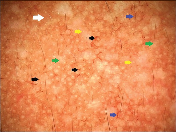
Figure 2.

Dermoscopy of the melasma lesion revealing diffuse light-to-dark brown (white arrow) pseudoreticular network, multiple brown dots, granules and globules (black arrows), arcuate and annular structures (blue arrows), with sparing of the perifollicular region (green arrows), and around the openings of sweat glands (yellow arrows) (polarizing mode, ×20)
