Figure 3.

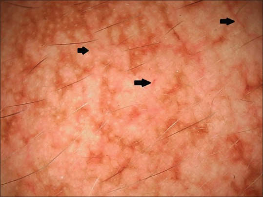
Dermoscopy of another melasma lesion revealing, in addition to the features seen in Figure 2, increased vascularity and telangiectasias (black arrows) (polarizing mode, ×20)

Dermoscopy of another melasma lesion revealing, in addition to the features seen in Figure 2, increased vascularity and telangiectasias (black arrows) (polarizing mode, ×20)