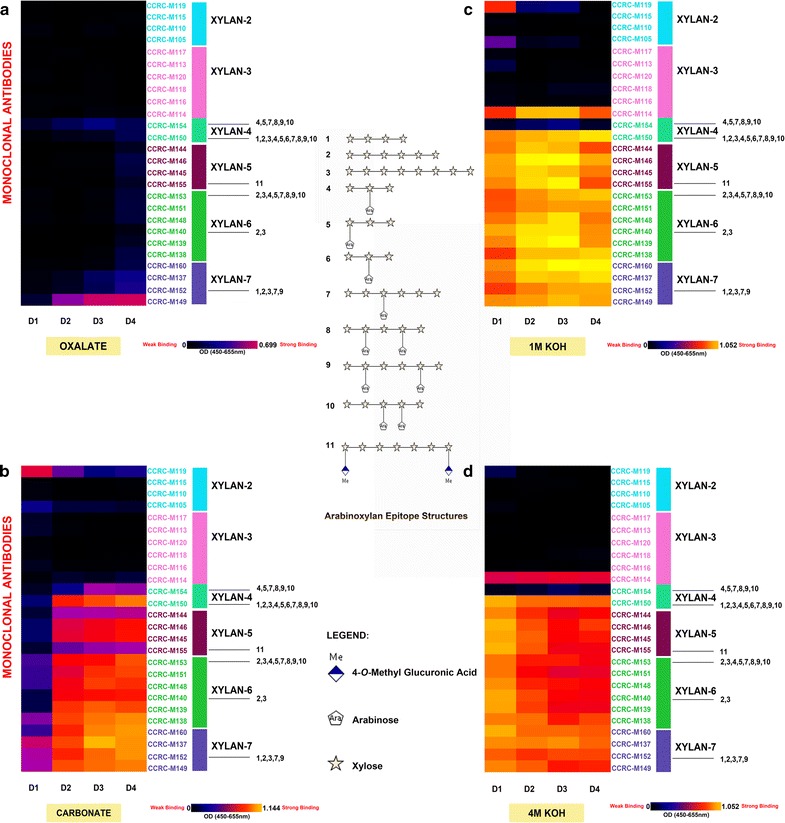
Fig. 2.

Xylan profiling of Col-0 inflorescence stems. ELISA binding signals specific to xylan epitope groups (Xylan2 to Xylan7) were isolated from this figure to depict distinct xylan epitopes enriched from different chemical extracts (a oxalate; b carbonate; c 1M KOH; d 4M KOH) with increasing harshness and at different stages (D1-D4) of Arabidopsis stem development. The ELISA heat map depicts signal binding strength where yellow, red, and black colours represent strong, medium, and no binding, respectively. The groups of mAbs are based on their specificity to different xylans at the right hand side of the figure. The top bar graph displays the mg soluble (glucose equivalent) recovered per gram of biomass. The middle illustration depicts the specific xylan epitope structures that xylan-directed specific mAbs bind to. Xylan epitope characterisation was based on the results of Schmidt et al. [6]
