Fig. 2.

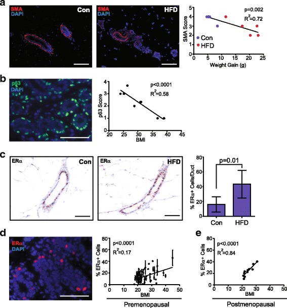
Obesity reduces basal/myoepithelial cells and enhances estrogen receptor (ER)α-positive luminal cells. a Basal/myoepithelial cells were detected using immunofluorescence (IF) for alpha smooth muscle actin (SMA, red) within the mammary glands of mice fed a control diet (Con) or high-fat diet (HFD). Nuclei were detected with 4′6-diamidine-2′-phenylidole dihydrochloride (DAPI) (blue). SMA continuity was scored as described in “Methods” and graphed in relation to weight gain. b p63+ (green) basal/myoepithelial cells were detected using IF within breast tissue from reduction mammoplasty surgery in women aged 20–45 years. Nuclei were detected with DAPI (blue). p63 expression was scored as described in “Methods” and graphed in relation to body mass index (BMI; n = 7). c ERα expression was immunohistochemically quantified within mammary glands from mice fed Con or HFD (5 mice/group; mean ± s.d.). d ERα expression (red) was detected using IF within human breast tissue from women ages 20–45 years. Nuclei were detected using DAPI (blue). ERα expression was plotted in relation to BMI (n = 39; mean ± s.d.). e ERα expression was plotted in relation to BMI, as reported through the Susan G. Komen Tissue Repository (n = 11). Magnification bar = 100 μm
